Ten organisations account for half of all animal research in Great Britain in 2021
-
99% of procedures carried out in mice, fish, and rats
-
83% of procedures caused similar pain (or less) than an injection
-
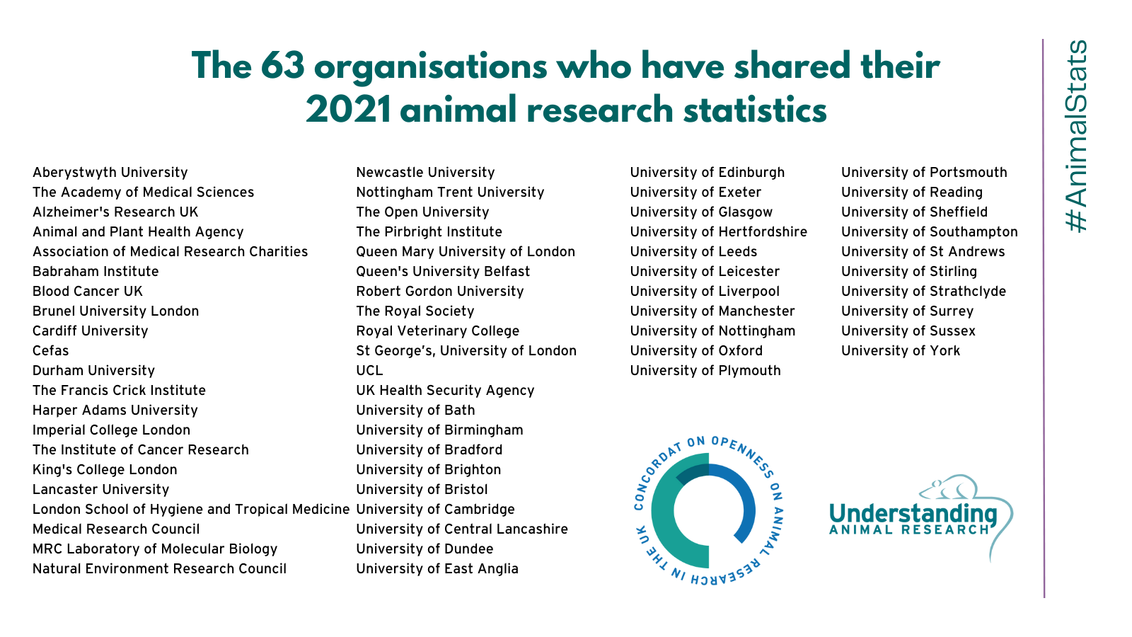
63 research institutions proactively share their 2021 animal research statistics
Today, 30 June 2022, Understanding Animal Research (UAR) has published a list of the ten organisations that carry out the highest number of animal procedures – those used in medical, veterinary, and scientific research – in Great Britain. These statistics are freely available on the organisations’ websites as part of their ongoing commitment to transparency and openness around the use of animals in research.
This list coincides with the publication of the Home Office’s report on the statistics of scientific procedures on living animals in Great Britain in 2021.
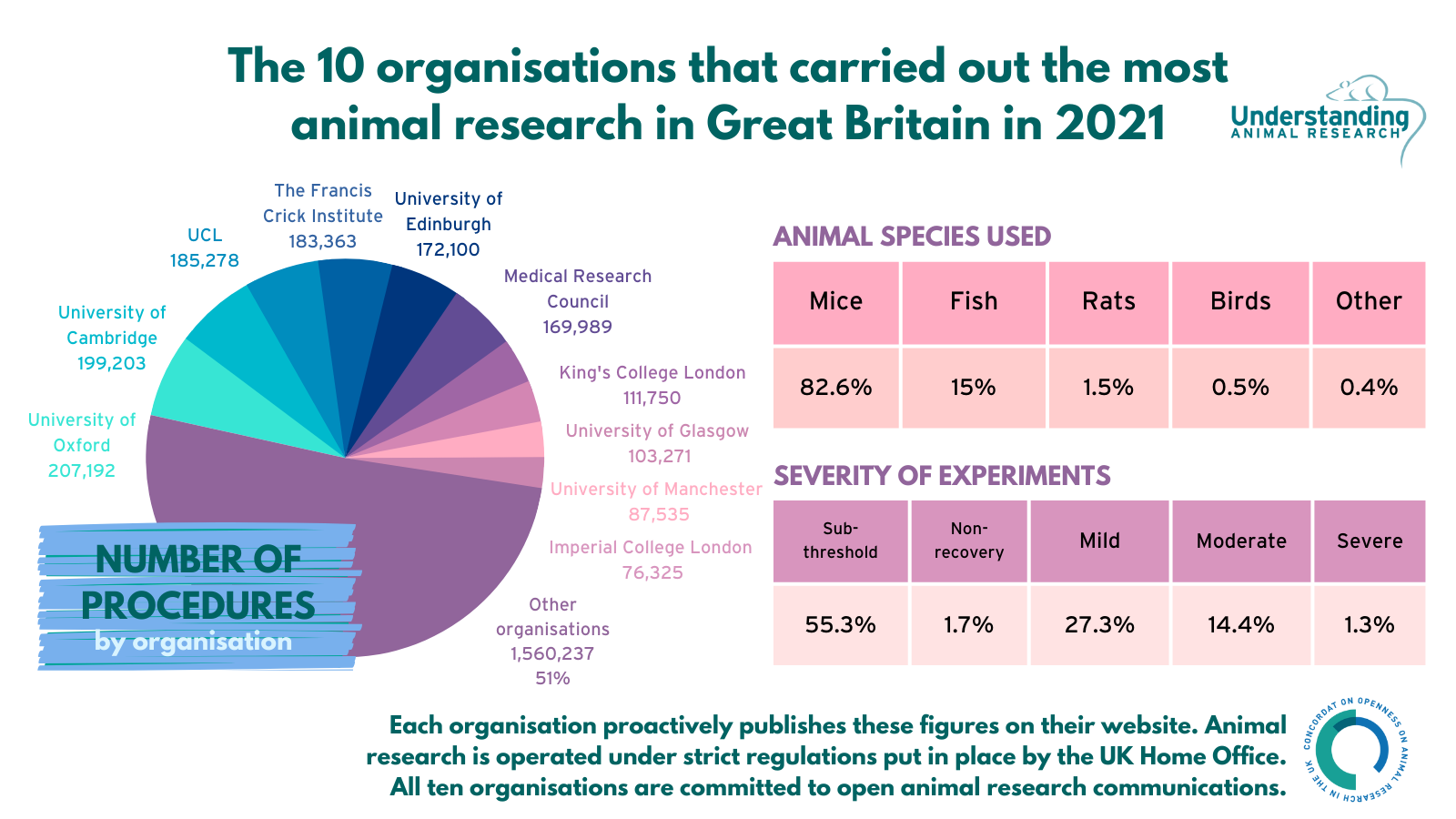
These ten organisations carried out 1,496,006 procedures, 49% or nearly half of the 3,056,243 procedures carried out on animals for scientific research in Great Britain in 2021*. Of these 1,496,006 procedures, more than 99% were carried out on mice, fish and rats and 83% were classified as causing a similar level of pain, or less, as an injection.
For more information on the 2021 animal research statistics for Great Britain, you can read our analysis.
The ten organisations are listed below alongside the total number of procedures they carried out in 2021. Each organisation’s name links to its animal research webpage, which includes more detailed statistics. This is the seventh consecutive year that organisations have come together to publicise their collective statistics and examples of their research.
|
Organisation |
Number of Procedures (2021) |
|
207,192 |
|
|
199,203 |
|
|
185,278 |
|
|
183,363 |
|
|
172,100 |
|
|
169,989 |
|
|
111,750 |
|
|
103,271 |
|
|
87,535 |
|
|
76,325 |
|
|
TOTAL |
1,496,006 |
63 organisations have published their 2021 animal research statistics
UAR has also produced a list (see appendix) of 63 organisations in the UK that have publicly shared their 2021 animal research statistics. This includes organisations that carry out and/or fund animal research.
All organisations are committed to the ‘3Rs’ of replacement, reduction and refinement. This means avoiding or replacing the use of animals where possible; minimising the number of animals used per experiment and optimising the experience of the animals to improve animal welfare. However, as institutions expand and conduct more research, the total number of animals used can rise even if fewer animals are used per study.
All organisations listed are signatories to the Concordat on Openness on Animal Research in the UK, a commitment to be more open about the use of animals in scientific, medical and veterinary research in the UK. More than 125 organisations have signed the Concordat including UK universities, medical research charities, research funders, learned societies and commercial research organisations.
Wendy Jarrett, Chief Executive of Understanding Animal Research, which developed the Concordat on Openness, said:
“Animal research remains a small but vital part of the quest for new medicines, vaccines and treatments for humans and animals. We know that the majority of the British public accepts that animals are needed for this research, but it is important that organisations that use animals in research maintain the public’s trust in them. By providing this level of information about the numbers of animals used, and the experience of those animals, as well as details of the medical breakthroughs that derive from this research, these Concordat signatories are helping the public to make up their own minds about how they feel about the use of animals in scientific research in Great Britain.”
Professor Anne Ferguson-Smith, Pro Vice-Chancellor for Research at the University of Cambridge:
“Animal research continues to be an important part of biomedical science, but as research institutions it is vital that we do not take public support for granted, and instead explain clearly why and how we work with animals and the steps we take to ensure good animal welfare.
“Since first signing the Concordat in 2014, Cambridge University has strived to be as open about our animal research as possible, sharing a wealth of information and case studies, and continuing to engage the public. We believe it’s important to show leadership in this area and we hope our efforts make a difference and show others within the sector what can be achieved.”
Professor David Lomas, UCL Vice-Provost (Health)
“Research using animals plays a small but vital role in UCL’s biomedical research, enabling us to make ground-breaking, life-saving advances; this is even more clear than ever as animal research enabled scientists to rapidly develop effective treatments and vaccines for Covid-19. Here at UCL we make every effort to ensure that animals are only used in research when strictly necessary, to treat them with utmost care, and to improve methods to minimise harm and maximise public benefit.”
Jan-Bas Prins, Director of the Francis Crick Institute’s Biological Research Facility:
“The number of procedures carried out at the Crick has remained fairly steady from 2020 to 2021 and they are down from years before the pandemic. The Crick is committed to the 3Rs and to providing a research environment where the development of and access to non-animal methods is a matter of course.”
Professor Jonathan Seckl, Senior Vice Principal at the University of Edinburgh:
"At the University of Edinburgh, we tackle some of the most difficult problems in human and animal health and the sustainability of our planetary ecosystems. We use animals for research where no alternatives exist. In the past year we have continued to make strides in the reduction, refinement and replacement of the use of animals in research. For example, we have developed 3D stem cell models to reduce the need for live birds for studies into common infections in chickens, which are a source of human diseases such as salmonella and ‘flu, as well as harming chickens and other birds themselves. Nevertheless, for the time being, the use of animals in research remains vital for developing new treatments for serious diseases of humans and other animals. The figures released today reflect the significant impact of Edinburgh and other UK biomedical research universities in addressing and solving some of the world’s greatest health challenges."
Claire Newland, MRC Director of Policy, Ethics and Governance:
“The MRC supports the highest quality research that has led to the development of life-saving treatments and advanced our understanding of basic human biology. The use of animals has been necessary for many of these discoveries, including in the development of next generation vaccines and other treatments essential to mitigating Covid-19 and other potential future pandemics. MRC is committed to ensuring the highest possible levels of animal welfare and to replacing, refining and reducing the use of animals where possible.”
Stephen Woodley, Director of King’s College London Biological Services:
“The use of animals in science is a small but important part of King’s College London's research into human disease and biomedical sciences. King’s College London is committed to ensuring the highest standards of animal welfare and ensuring the 3Rs are adhered to. As part of our commitment to 3Rs, the number of animals used in research at King's College London is kept to a minimum, while still allowing for scientifically valid data.”
David Duncan, University of Glasgow Deputy Vice Chancellor and Chief Operating Officer:
“Research using animals makes a vital contribution to the understanding, treatment and cure of a range of major diseases in humans, such as cancer and Alzheimer’s. Animals are used in research only where it is essential. While the University is committed to the development of alternative methods – such as computer modelling, tissue culture, cell and molecular biology, and research with human material – some work involving animals must continue for further advances to be made. The University is committed to the principles of reduction, refinement, and replacement. All research undertaken on animals is conducted under strict ethical and welfare guidelines, under licence by the Home Office.”
Maria Kamper, Director of the Animal Research Unit at The University of Manchester:
“We are proud to be one of the first UK institutions to embrace openness and transparency about our animal research. A virtual tour, facts, figures, project summaries and case studies, and lots more besides are freely available on our website.
“We are also proud of the high ethical standards with which we carry out our work and the way we care for our animals. Though we are strongly committed to replacing animals wherever possible with alternatives, reducing their numbers and refining the work we do to ever improve their welfare, animals still play a hugely important role in scientific research.
“Our research involving animals helps us understand how biological systems work so we can find ways to treat disease and understand not just human – but also animal health. That is why animal research remains a critical way for scientists to develop of new medicines and cutting-edge medical technologies.”
Professor Marina Botto, Director of Bioservices, Imperial College London
“Imperial’s commitment to the 3Rs principles and openness around animal research is reflected in our animal numbers. Research using animals enables us to understand human diseases and how they can be treated. We are also committed to the highest standard of animal welfare as demonstrated by the AAALAC accreditation”
-Ends-
Notes to Editors
For more information, contact Hannah Hobson on 07759235176 or hhobson@uar.org.uk.
The hashtag for social media is #AnimalStats.
Understanding Animal Research (UAR) is a not-for-profit organisation that explains how and why animals are used in scientific research in the UK. UAR promotes open communications about animal research.
A list of recent animal research case studies from contributing organisations can be found below.
Further information on the Concordat on Openness on Animal Research in the UK can be found here: http://concordatopenness.org.uk
These figures refer to procedures using animals for medical, veterinary, or scientific research, as licensed by the UK’s Home Office under the Animals (Scientific Procedures) Act 1986. The use of animals to test tobacco products was banned in the UK in 1997 and it has been illegal to use animals to test cosmetic products in this country since 1998. A policy ban on household product testing using animals was introduced in 2010. Since 2013, it has been illegal to sell or import cosmetics anywhere in the UK or the EU where the finished product or its ingredients have been tested on animals.
*The Home Office recorded 3,056,243 completed procedures in 2021, 1,496,006 (49%) of which were carried out at these ten organisations.
Full table of procedures broken down by species from top ten organisations
|
Organisation |
Total Procedures (2021) |
Mice |
Fish |
Rats |
Birds |
Non-Human Primates |
Other |
|
207,192 |
197,908 |
7,848 |
1,188 |
|
13 |
235 |
|
|
199,203 |
152,471 |
43,850 |
2,095 |
|
33 |
754 |
|
|
185,278 |
112,925 |
69,269 |
2,562 |
|
|
522 |
|
|
183,363 |
166,827 |
16,295 |
|
|
|
241 |
|
|
172,100 |
111,397 |
45,462 |
8,104 |
6,132 |
|
1,005 |
|
|
169,989 |
169,920 |
|
12 |
|
57 |
|
|
|
111,750 |
89,258 |
19,574 |
2,599 |
|
|
638 |
|
|
103,271 |
96,784 |
3,775 |
1,554 |
380 |
|
778 |
|
|
87,535 |
70,586 |
13,328 |
2,646 |
|
|
975 |
|
|
76,325 |
68,085 |
5,528 |
1,631 |
239 |
|
842 |
|
|
TOTAL |
1,496,006 |
1,236,161 (82.6%) |
224,929 (15.0%) |
22,391 (1.5%) |
6,751 (0.5%) |
103 (0.01%) |
5,990 (0.4%) |
All numbers represent completed procedures on animals in 2021. The number of procedures carried out using animals will be slightly higher than the number of animals used, as a small number of animals may be used in more than one procedure.
Full table of procedures broken down by severity categories from top ten organisations
|
Organisation |
Sub-Threshold |
Non-Recovery |
Mild |
Moderate |
Severe |
Total Procedures (2021) |
|
132,216 |
3,433 |
38,050 |
31,613 |
1,880 |
207,192 |
|
|
81,569 |
1,041 |
87,971 |
26,258 |
2,364 |
199,203 |
|
|
55,718 |
11,062 |
94,393 |
23,336 |
769 |
185,278 |
|
|
128,238 |
262 |
23,096 |
28,159 |
3,608 |
183,363 |
|
|
117,190 |
2,741 |
31,513 |
17,949 |
2,707 |
172,100 |
|
|
115,434 |
1,351 |
40,719 |
10,532 |
1,953 |
169,989 |
|
|
64,203 |
3,023 |
22,408 |
19,337 |
2,779 |
111,750 |
|
|
62,180 |
758 |
17,699 |
20,983 |
1,651 |
103,271 |
|
|
48,252 |
526 |
17,995 |
19,875 |
801 |
87,449 |
|
|
22,602 |
1,732 |
34,705 |
16,772 |
514 |
76,325 |
|
|
TOTAL |
827,602 (55.3%) |
25,929 (1.7%) |
408,549 (27.3%) |
214,814 (14.4%) |
19,026 (1.3%) |
1,495,920 |
The University of Manchester carried out 86 procedures on animals not licensed under the Animals (Scientific Procedures) Act 1986, These 86 procedures are not rated with a severity band and are therefore not included in the above table.
Examples of severity
Severity assessments measure the harm experienced by an animal during a procedure. A procedure can be as mild as an injection, or as severe as an organ transplant. Severity assessments reflect the peak severity of the entire procedure and are classified into five different categories:
Sub-threshold: When a procedure did not cause suffering above the threshold for regulation, i.e. it was less than the level of pain, suffering, distress or lasting harm that is caused by inserting a hypodermic needle according to good veterinary practice.
Non-recovery: When the entire procedure takes place under general anaesthetic and the animal is humanely killed before waking up.
Mild: Any pain or suffering experienced was only slight or transitory and minor so that the animal returns to its normal state within a short period of time. For example, the equivalent of an injection or having a blood sample taken.
Moderate: The procedure caused a significant and easily detectable disturbance to an animal’s normal state, but this was not life threatening. For example, surgery carried out under general anaesthesia followed by painkillers during recovery.
Severe: The procedure caused a major departure from the animal’s usual state of health and well-being. This would usually include long-term disease processes where assistance with normal activities such as feeding and drinking were required, or where significant deficits in behaviours/activities persist. Animals found dead are commonly classified as severe as pre-mortality suffering often cannot be assessed.
List of 63 UK organisations that have shared their 2021 animal research statistics:
- Aberystwyth University
- The Academy of Medical Sciences
- Alzheimer's Research UK
- Animal and Plant Health Agency
- Association of Medical Research Charities
- Babraham Institute
- Blood Cancer UK
- Brunel University London
- Cardiff University
- Cefas
- Durham University
- The Francis Crick Institute
- Harper Adams University
- Imperial College London
- The Institute of Cancer Research
- King's College London
- Lancaster University
- London School of Hygiene and Tropical Medicine
- Medical Research Council
- MRC Laboratory of Molecular Biology
- Natural Environment Research Council
- Newcastle University
- Nottingham Trent University
- The Open University
- The Pirbright Institute
- Queen Mary University of London
- Queen's University Belfast
- Robert Gordon University
- The Royal Society
- Royal Veterinary College
- St George’s, University of London
- UCL
- UK Health Security Agency
- University of Bath
- University of Birmingham
- University of Bradford
- University of Brighton
- University of Bristol
- University of Cambridge
- University of Central Lancashire
- University of Dundee
- University of East Anglia
- University of Edinburgh
- University of Exeter
- University of Glasgow
- University of Hertfordshire
- University of Leeds
- University of Leicester
- University of Liverpool
- University of Manchester
- University of Nottingham
- University of Oxford
- University of Plymouth
- University of Portsmouth
- University of Reading
- University of Sheffield
- University of Southampton
- University of St Andrews
- University of Stirling
- University of Strathclyde
- University of Surrey
- University of Sussex
- University of York
CASE STUDIES
University of Oxford
New research sheds light on how ultrasound could be used to treat psychiatric disorders
A new study in macaque monkeys has shed light on which parts of the brain support credit assignment processes (how the brain links outcomes with its decisions) and, for the first time, how low-intensity transcranial ultrasound stimulation (TUS) can modulate both brain activity and behaviours related to these decision-making and learning processes.
While currently developed in an animal model, although in a brain area homologous to the one in humans, this line of research and the use of TUS could one day be applied to clinical research to tackle psychiatric conditions where maladaptive decisions are observed.
The study published in the journal Science Advances shows that credit assignment-related activity in this small lateral prefrontal area of the brain, which supports adaptive behaviours, can be safely, reversibly and quickly disrupted with TUS.
After stimulating this brain area, the animals in the study became more exploratory in their decisions. As a consequence of the ultrasound neuromodulation, behaviour was no longer guided by choice value – meaning that they could not understand that some choices would cause better outcomes – and decision-making was less adaptive in the task.
The study also showed that this process remained intact if another brain region (also part of the prefrontal cortex) was stimulated as control condition; showing for the first time how task-related brain modulation is specific to stimulation of specific areas that mediate a certain cognitive process.
The first author, Dr Davide Folloni of Oxford’s Wellcome Centre for Integrative Neuroimaging, said: 'This research has critical importance in a number of areas, including allowing us for the first time to non-invasively test hypothesis on the role of deep cortical areas in cognition while simultaneously recording the underlying neural activity in primates and potentially humans. This could significantly improve clinical treatment by helping surgeons to test implant sites for suitability before surgery, greatly improving the efficiency and accuracy of such delicate surgery. By improving our knowledge of the contribution of previously inaccessible dysfunctional brain areas in psychiatric and neurological diseases this will also open up new avenues for non-invasive treatment for a number or neurological conditions.'
University of Cambridge
Scientists reverse age-related memory loss in mice
Scientists at Cambridge and Leeds have successfully reversed age-related memory loss in mice and say their discovery could lead to the development of treatments to prevent memory loss in people as they age.
In a study published in Molecular Psychiatry, the team show that changes in the extracellular matrix of the brain – ‘scaffolding’ around nerve cells – lead to loss of memory with ageing, but that it is possible to reverse these using genetic treatments.
Professor James Fawcett from the John van Geest Centre for Brain Repair at the University of Cambridge said: “What is exciting about this is that although our study was only in mice, the same mechanism should operate in humans – the molecules and structures in the human brain are the same as those in rodents. This suggests that it may be possible to prevent humans from developing memory loss in old age.”
The team have already identified a potential drug, licensed for human use, which can be taken by mouth and inhibits the formation of perineuronal nets (PNNs), which are connected with age-related memory decline. When this compound is given to mice and rats it can restore memory in ageing and also improves recovery in spinal cord injury. The researchers are investigating whether it might help alleviate memory loss in animal models of Alzheimer's disease.
https://www.cam.ac.uk/research/news/scientists-reverse-age-related-memory-loss-in-mice
UCL
Magnetic seeds used to heat and kill cancer
Scientists at UCL have developed a novel cancer therapy, now demonstrated in mice, that uses an MRI scanner to guide a magnetic seed through the brain to heat and destroy tumours.
The therapy is called “minimally invasive image-guided ablation” or MINIMA and comprises a ferromagnetic thermoseed navigated to a tumour using magnetic propulsion gradients generated by an MRI scanner, before being remotely heated to kill nearby cancer cells.
Researchers say the findings, published in Advanced Science, establish ‘proof-of-concept’ for precise and effective treatment of hard-to-reach glioblastoma, along with other cancers such as prostate, that could benefit from less invasive therapies. They say the therapy has the potential to extend survival, reduce recovery times, and to avoid traditional side effects by precisely treating the tumour without harming healthy tissues. Because the heating seed is magnetic, the magnetic fields in the MRI scanner can be used to remotely steer the seed through tissue to the tumour. Once at the tumour, the seed can then be heated, destroying the cancer cells, while causing limited damage to surrounding healthy tissues.
In the study, the UCL Centre for Advanced Biomedical Imaging team demonstrate the three key components of MINIMA to a high level of accuracy: precise seed imaging; navigation through brain tissue using a tailored MRI system, tracked to within 0.3 mm accuracy; and eradicating the tumour by heating it in a mouse model.
Ferromagnetic thermoseeds are spherical in shape, 2 mm in size and are made of a metal alloy; they are implanted superficially into tissue before being navigated to the cancer.
MRI scanners are readily available in hospitals around the world and are pivotal in the diagnosis of diseases such as cancer. The work at UCL shows that MINIMA has the potential to elevate an MRI scanner from a diagnostic device to a therapeutic platform.
https://www.ucl.ac.uk/news/2022/feb/magnetic-seeds-used-heat-and-kill-cancer
The Francis Crick Institute
Gene-editing used to create single sex mice litters
Scientists at the Francis Crick Institute, in collaboration with University of Kent, have used gene editing technology to create female-only and male-only mice litters with 100% efficiency.
This proof of principle study, published in Nature Communications, demonstrates how the technology could be used to improve animal welfare in scientific research and agriculture.
In scientific research and farming, there is often a need for either male or female animals. For example, laboratory research into male or female reproduction requires only animals of the sex being studied. And in farming, only female animals are required for egg production and in dairy herds. This means it is common practice for animals of the unrequired sex to be culled after birth.
The researchers’ new method uses a two-part genetic system to inactivate embryos shortly after fertilisation, allowing only the desired sex to develop. Such a genetically-based method to control the sex of offspring could drastically reduce culling in both industries.
https://www.crick.ac.uk/news/2021-12-03_gene-editing-used-to-create-single-sex-mice-litters
University of Edinburgh
Drug firm licenses cancer discovery developed in mice
A new drug compound, discovered using studies with mice, is set to be transformed into medicines for hard-to-treat cancers.
The University of Edinburgh has signed a licensing deal with a US biopharmaceutical company, granting them exclusive worldwide rights to develop and commercialise treatments based on the compound, known as NXP900.
NXP900 works by blocking proteins called SRC and YES1, which have been linked to several types of cancer. In cell and animal studies, NXP900 has shown the potential to reduce the growth of certain types of breast, colon, prostate, pancreatic and ovarian cancer, as well as tumours affecting the lungs, head and neck and oesophagus.
Its discovery follows 10 years of research at the Cancer Research UK Edinburgh Centre within the University of Edinburgh’s Institute of Genetics and Cancer.
https://www.ed.ac.uk/research/animal-research/news/drug-firm-licenses-cancer-discovery
Medical Research Council
Insights into mitochondrial disorders from animal research
The use of animals in fundamental biomedical research helps researchers better understand the biological processes that are central to our health. This is essential for developing safe and effective ways of preventing or treating disease. Defective mitochondria – the ‘batteries’ that power the cells of our bodies – could in future be repaired using gene-editing techniques. Scientists at the MRC Mitochondrial Biology Unit at the University of Cambridge have shown that it is possible to modify the mitochondrial genome in live mice, paving the way for new treatments for incurable mitochondrial disorders.
Our cells contain mitochondria, which provide the energy for our cells to function. Each of these mitochondria contains a tiny amount of mitochondrial DNA. Mitochondrial DNA makes up only 0.1% of the overall human genome and is passed down exclusively from mother to child.
Faults in our mitochondrial DNA can affect how well the mitochondria operate, leading to mitochondrial diseases. These diseases are serious and often fatal conditions that affect around 1 in 5,000 people. They are incurable and largely untreatable.
There are typically around 1,000 copies of mitochondrial DNA in each cell, and the percentage of these that are damaged, or mutated, will determine whether a person will suffer from mitochondrial disease or not. Usually, more than 60% of the mitochondria in a cell need to be faulty for the disease to emerge, and the more defective mitochondria a person has, the more severe their disease will be. If the percentage of defective DNA could be reduced, the disease could potentially be treated.
In 2018, a team from the MRC Mitochondrial Biology Unit at the University of Cambridge applied an experimental gene therapy treatment in mice and were able to successfully target and eliminate the damaged mitochondrial DNA in cells, allowing mitochondria with healthy DNA within that cell to take their place. However, this approach is only possible in cells that contain a mixture of healthy and faulty mitochondrial DNA. There has to be enough healthy mitochondrial DNA in the cell so it can copy itself and replace the faulty ones that had been removed. It would not work in cells whose entire mitochondria had faulty DNA.
To tackle this limitation, the team used a biological tool known as a mitochondrial base editor to edit the mitochondrial DNA of live mice. The result, published in February 2022, is a proof-of-concept study that shows how it is indeed possible to edit the mitochondrial DNA in mice. The treatment is delivered into the bloodstream of the mouse using a modified virus, which is then taken up by its cells. The tool looks for a unique sequence of base pairs – combinations of the A, C, G and T molecules that make up DNA. It then changes the DNA base – in this case, changing a C to a T. This would, in principle, enable the tool to correct certain ‘spelling mistakes’ that cause the mitochondria to malfunction.
There are currently no suitable mouse models of mitochondrial DNA diseases, so the researchers used healthy mice to test the mitochondrial base editors. However, it shows that it is possible to edit mitochondrial DNA genes in a live animal.
This ground-breaking research is the first time that researchers have been able to change DNA base pairs in mitochondria in a live animal. It shows that, in principle, scientists can go in and correct spelling mistakes in defective mitochondrial DNA, producing healthy mitochondria that allow the cells to function properly.
King’s College London
Scientists obtain first high-resolution 3D image of muscle protein
Heart and skeletal muscle owe their function as reliable biological machines to the extraordinary precision with which their smallest contractile structures, the sarcomeres, are assembled. Generating the power to deliver blood to all organs and enabling body movements – from breathing to sprint running – these muscles require the coordinated assembly of millions of protein components which are coordinated by the largest proteins of the human body, the giant ‘rulers’: titin, nebulin and obscurin.
Due to their large size, studying these proteins is fraught with technical challenges. Scientists have now used cutting-edge electron microscopy techniques to obtain the first high-resolution 3D image of nebulin, a giant actin-binding protein that is an essential component of skeletal muscle. This discovery allows us an opportunity to better understand the role of nebulin, as its functions have remained vague due to its large size and the difficulty in extracting nebulin in a native state from muscle.
Professor Mathias Gautel, Head of the School of Basic & Medical Biosciences, Dr Ay Lin Kho and a team of Max Planck researchers led by Stefan Raunser, used electron cryo-tomography to decipher the structure of nebulin in impressive detail. Their findings could lead to novel therapeutic approaches to treat muscular diseases, as genetic mutations in nebulin are accompanied by a dramatic loss in muscle force known as nemaline myopathy.
Skeletal and heart muscles contract and relax upon sliding of parallel filaments of the proteins myosin and actin. Nebulin, another long slender protein, which is present only in skeletal muscle, pairs up with actin, stabilising and regulating it by a series of similar structures called repeats. Mutations in the gene encoding nebulin can produce an abnormal nebulin that causes nemaline myopathy, an incurable neuromuscular disorder with various degrees of severity, from muscle weakness to speech impediments and respiratory problems.
Knowing the structure of nebulin and how exactly it interacts with actin could be pivotal to the development of new treatments. In this study, the researchers were able visualise myosin and actin directly in their native environment, the muscle, by using a powerful microscopy technique called electron cryo-tomography (cryo-ET). Experiments were done on mouse muscles that are very similar to human ones, using procedures optimised in labs at King’s College London’s Randall Centre for Cell & Molecular Biophysics. Strikingly, heart muscle actin filaments demonstrated the clear absence of nebulin.
The findings reveal that each nebulin repeat binds with an actin subunit, demonstrating nebulin’s role as a ruler that gradates the length of actin filaments. Each nebulin repeat interacts with every neighbouring actin subunit, which explains its role as a stabiliser. Finally, the scientists propose that nebulin could regulate the binding of actin and myosin, and hence muscle contraction, by interacting with another protein called troponin.
Driven by their recent success, the group will now concentrate on unveiling the structural details of myosin regulation, the motor filament of muscle. Such findings could finally help paint the complete picture of the intricate details behind skeletal and heart muscle contraction.
https://www.kcl.ac.uk/news/scientists-obtain-first-high-resolution-3d-image-of-muscle-protein
University of Glasgow
The University of Glasgow, along with collaborators at the Beatson Institute for Cancer Research, have a world-leading reputation for translational cancer research. A recent study, published in Cell Death and Differentiation and led by Prof Stephen Tait, investigated targeting glioblastoma tumour cells – the most common form of brain cancer, that has few therapeutic options – with new drugs called BH3-mimetics, which directly target cell death. By using mouse models alongside patient material, the study demonstrated that two types of BHS-mimetics are particularly effective in glioblastoma and, importantly, by applying them sequentially, showed improved survival in mouse models without toxic side effects. The team behind the study hopes to explore these drugs further, as a potential new therapeutic approach for glioblastoma.
University of Manchester
Sex drug can strongly suppress abnormal heart rhythms, finds study
The drug Viagra, used to treat sexual impotence, can strongly suppress abnormal heart rhythms known as arrythmias in sheep according to University of Manchester scientists. The British Heart Foundation funded study could have important implications on the management of the condition on humans.
In female sheep, the drug - also known as sildenafil - was able to suppress an arrythmia called Torsades de Pointes within 90 seconds by reducing the frequency of irregular heart rhythms caused by abnormal handling of calcium. It also reduced the probability of Torsades de Pointes, which can lead to sudden cardiac death. However, the team believe the drug could treat other arrythmias as well.
Cardiac arrythmias often follow a heart attack or result from heart disease. Though often benign, they can cause symptoms including palpitations, breathlessness, fainting and sometimes sudden death. Treatments such as beta blockers and implantable defibrillators exist, though are not always effective.
Working on isolated sheep heart muscle cells known as myocytes, the research team were also able to measure their calcium stores, as well as their electrical properties. Although calcium is a key driver of the heart’s pumping action, its overload can be a root cause of arrythmias. Viagra was able to suppress the mechanism in the cell which causes calcium overload.
Imperial College London
Metal-based molecules show promise against the build-up of Alzheimer’s peptides
In lab tests, Imperial researchers have created a metal-based molecule that inhibits the build-up of a peptide associated with Alzheimer’s disease.
A peptide is a fragment of a protein, and one of the key hallmarks of Alzheimer's disease is the build-up of a specific peptide known as amyloid-β. The team demonstrated that with the aid of ultrasound, their molecule can cross the blood-brain barrier in mice, targeting the part of the brain where the damaging peptide most often accumulates.
Alzheimer's disease is the most common form of dementia, affecting approximately 50 million people worldwide. There is a pressing need to develop drugs that can prevent or reverse the effects of this devastating disease.
Some metal-based molecules have been previously designed to prevent amyloid-β from building up. However, these are often toxic to cells, or are unable to cross the blood-brain barrier (BBB) – a semi-permeable protective barrier that carefully regulates the passage of substances that enter and exit the brain.
Now, a team from the Departments of Chemistry and Bioengineering at Imperial College London have designed a metal-based molecule that is highly effective at preventing the build-up of amyloid-β in lab-based studies. They also showed that the molecule is non-toxic to human brain-like cells and that it can cross the blood-brain barrier in mice with the help of a technique using microbubbles and focused ultrasound. The results are reported in the journal Chemical Science.
https://www.imperial.ac.uk/news/226313/metal-based-molecules-show-promise-against-build-up/
Last edited: 1 November 2022 16:54
