Haemophilia is a rare, inherited bleeding disorder in which blood cannot clot normally. Long known as the "Royal Disease", haemophilia affected generations of royal families in England, Prussia and Russia, undoubtedly changing the course of history. Many male descendants of Queen Victoria were plagued with the disease that was transmitted by female members, hidden in their X chromosome.
A genetic disease
The disorder occurs because certain blood clotting factors are missing or do not work properly. Because a clot doesn’t form, extensive external or internal bleeding can occur which can eventually damage articulations and even cause death. The tiniest cut can become a gushing flow of blood.
There are two types of inherited haemophilia:
- Type A, the most common type, is caused by a deficiency of factor VIII, one of the proteins that helps blood to form clots.
- Type B haemophilia is caused by a deficiency of factor IX.
Although haemophilia is usually diagnosed at birth, the disorder can also be acquired later in life if the body begins to produce antibodies that attack and destroy clotting factors. However, this acquired type of haemophilia is very rare.
The main treatment for severe haemophilia involves receiving replacement of the specific clotting factor that you need, through a tube placed in a vein. This replacement therapy can be given to combat a bleeding episode that's in progress or on a regular schedule as preventative measures. Replacement clotting factor can be made from donated blood. Similar products, called recombinant clotting factors, aren't made from human blood. For some years, one option for treating people who have an inhibitor to factor VIII has been to give them porcine factor VIII. Porcine factor VIII is taken from the blood of pigs. The pig factor VIII is close enough to human factor VIII to stop bleeding but is different enough so that the inhibitors don't destroy it. Unfortunately, with repeated use, people can develop resistance against the porcine factor. Other forms of therapies include using hormones to stimulate the release of clotting factors or medication to help clots from breaking down and to stimulate their formation.
Why is research in animals still needed?
Although these therapies have been very successful, some challenging and unresolved tasks remain, such as reducing bleeding rates, the presence of joint damage, eliminating the development of inhibitors, and increasing the success rate of immune-tolerance induction (ITI).
Indeed, although very successful, patients tend to develop a resistance to clot proteins injected as treatment and inducing neutralising antibodies or “inhibitors”. The immune system recognises the factor concentrate as a foreign invader and develops inhibitors against the factor. Patients with inhibitors can no longer use FVIII/FIX concentrates and require more expensive bypass products or elimination of the inhibitors by ITI.
Many preclinical trials are carried out on animal models for haemophilia to tackle these challenges. Transnational research from haemophilia animal models gives valuable information about safety and efficacy, and guides design of human clinical trials. Animal models are also important for basic biological and preclinical studies. Suitable animal models are needed for greater advances in treating haemophilia, such as the development of better models for evaluation of the efficacy and safety of long-acting products, more powerful gene therapy vectors than are currently available, and successful ITI strategies. Mice, dogs, and pigs are the most commonly used animal models for haemophilia.
Animal models naturally affected - Dogs
The earliest recognised cases of haemophilia in dogs were documented in 1935 in three related Scottish terriers. Canine haemophilia A is essentially identical to the human disease in its clinical presentation characterised by severe-intermittent episodes of joint bleeding and haemorrhage.
Dogs with naturally occurring haemophilia A were first documented following observations of abnormally prolonged bleeding that could be treated or prevented by infusion of canine FVIII. Haemophilia A dogs have been used extensively in preclinical trials of human FVIII protein products as well as in studies on the safety and efficacy of gene therapy, and have provided promising data for bypass therapy.
Equally, researchers have identified at least three colonies of haemophilia B dogs, each of which has a unique molecular FIX defect. Similar to human haemophilia B, canine haemophilia B has a sex-linked inheritance pattern, and no detectable circulating FIX exists in the plasma. Translation data produced from haemophilia B dogs have supported the development of long-acting FIX with accompanying recent human clinical trials.
Sheep
Phenotypes consistent with human haemophilia A, including spontaneous bleeding and plasma FVIII activity of less than 1 %, have been found in sheep. Haemophilia sheep have been used in studies relevant to gene and cell therapies for haemophilia, including investigations of pre-existing immunity to AAV vectors and the use of mesenchymal stem cells as cellular delivery vehicles for the FVIII gene.
Rats
Researchers identified an inbred rat strain which tended to show abnormal haemorrhaging, a prolonged activated partial thromboplastin time, and a mutated FVIII gene. In rats, the FVIII gene is located on chromosome 18. Thus, haemophilia in rats is autosomal recessive, which contrasts with humans and other animal models where it is an X-linked hereditary disease. The mutated FVIII region of the rats generally lacks immunodominant epitopes for inhibitor formation thus this model may not be suitable for studying the immune response to FVIII treatment or developing ITI strategies.
Genetically engineered animal models - Mice
Although spontaneous bleeding does not naturally occur in mice, genome-editing technologies have contributed to development of various haemophiliac mouse models. Haemophilia mice are the best initial model to use when attempting to test new therapeutics because they only require small amounts of drugs.
In 1995, the first two haemophilia A mouse models were established. These two strains of FVIII knockout mice were generated through gene targeting and embryonic stem cell manipulation. Mice had no detectable circulating FVIII, and their plasma FVIII activity was less than 1 %. Unlike human haemophilia A, little spontaneous bleeding is observed in mouse models of haemophilia A, whereas tail clipping or other invasive procedures could lead to death. Mouse models of haemophilia A are widely used for the evaluation of FVIII treatment efficacy, investigation of mechanisms of inhibitor formation, and development of ITI protocols for FVIII.
Although mice with naturally occurring deficiency of FIX to mimic haemophilia B have not been identified, a series of FIX knockout mice were engineered using embryonic stem cells. Similar to haemophilia A mice, haemophilia B mice do not show spontaneous bleeding, but will bleed and die after tail clipping unless the wound is cauterised. Haemophilia B mice have been used to test the efficacy of FIX and FIX variants, including those FIX variants with very high clotting activities, as well as to evaluate the immunity and safety of gene therapy.
Researcher also modified the haemophilia A mouse model to be “humanised” for HLA class II antigen to understand the regulation of antibody responses against FVIII in haemophilia A. Use of this humanised haemophilia A mouse model allowed identification of peptides that trigger inhibitor formation, as well as the characterisation of interactions of T-cell receptors with disease-associated FVIII peptides.
CRISPR-Cas9 was also recently instrumental in constructing a reliable animal model for elucidating the humeral and cellular immune responses of patients to FVIII/FIX treatment. The CRISPR/Cas9 system and immunodeficient NSG mice were combined to mutate the FVIII and FIX genes, generating haemophilia A/B mice particularly useful for studying the human immune response to therapeutics.
Pigs
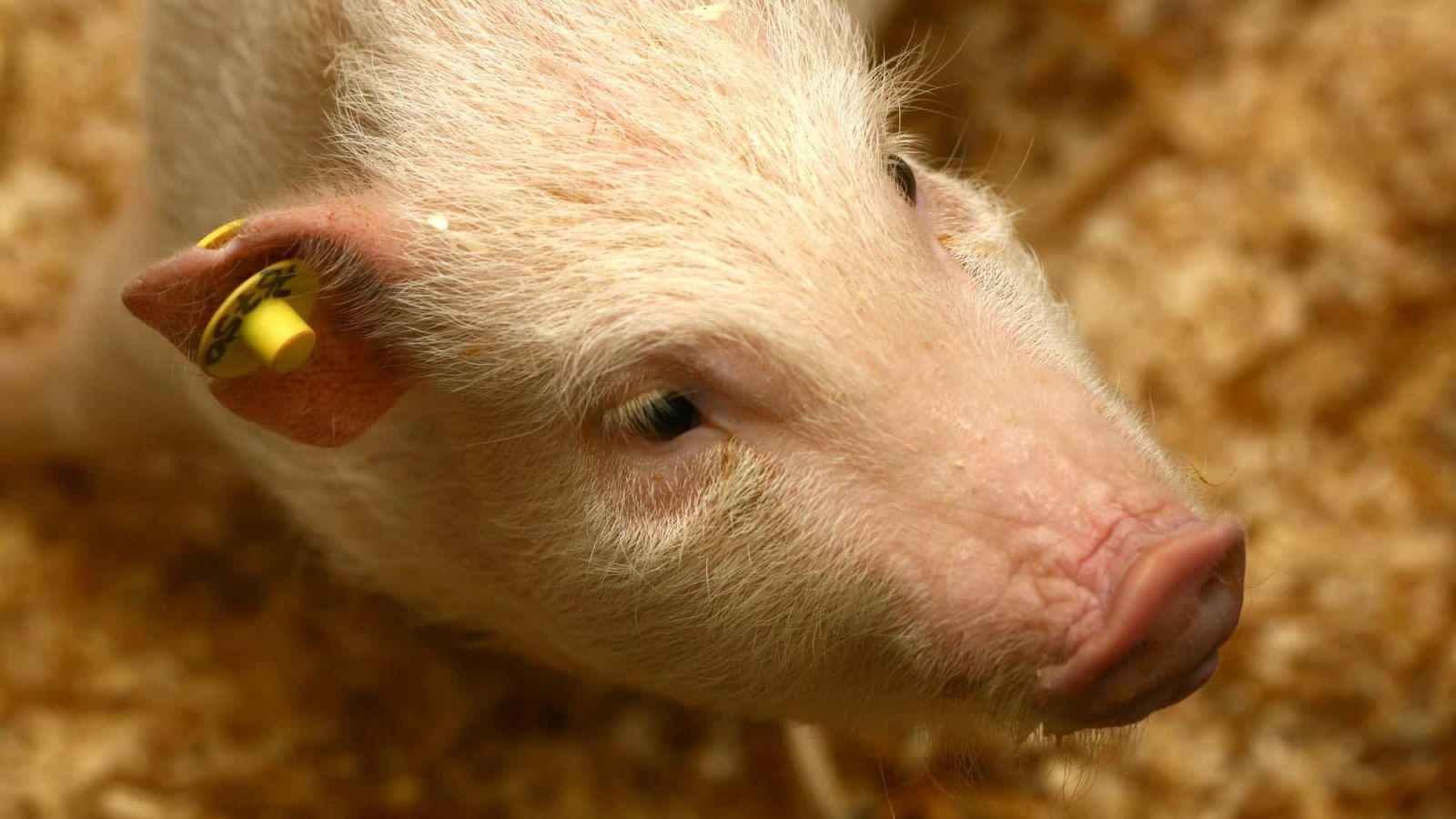
Pigs with haemophilia A were generated by nuclear transfer and cloning from porcine fetal fibroblasts. Species differences between humans and mice, such as size, general physiology, anatomy, and lifespan, limit the value of mouse models in preclinical trials. In contrast, the coagulation systems of pigs and humans are highly homologous. Moreover, haemophilia A pigs may develop arthropathy similar to humans because of repeated joint bleeding. Thus, haemophilia A pigs provide another option for evaluating novel therapeutics for hemophilia A patients. The haemophilia A pig is a severe hemophilia A animal model for studying not only haemophilia A gene therapy but also the next generation recombinant coagulation factors, such as recombinant factor VIII variants with a slower clearance rate.
For over 15 years, scientists have also worked with transgenic pigs that have been genetically engineered to produce human factor proteins in their milk.
REFERENCES
https://www.mayoclinic.org/diseases-conditions/hemophilia/diagnosis-treatment/drc-20373333
https://journals.plos.org/plosone/article?id=10.1371/journal.pone.0049450
https://www.hog.org/publications/detail/hogs-help-people-with-hemophilia-again
https://www.scientificamerican.com/article/in-the-fight-against-haemophilia-dogs-are-a-weapon/
https://www.ncbi.nlm.nih.gov/pubmed/26924758
https://www.ncbi.nlm.nih.gov/pubmed/19109232
https://www.ncbi.nlm.nih.gov/pubmed/12515715
https://www.ncbi.nlm.nih.gov/pubmed/11545609
https://www.ncbi.nlm.nih.gov/pubmed/26149014
https://www.ncbi.nlm.nih.gov/pmc/articles/PMC5056469/
https://www.ncbi.nlm.nih.gov/pubmed/7647782
https://www.ncbi.nlm.nih.gov/pubmed/20059562
https://www.ncbi.nlm.nih.gov/pubmed/23502223
https://www.ncbi.nlm.nih.gov/pubmed/20539913
https://www.ncbi.nlm.nih.gov/pubmed/27028920
https://www.ncbi.nlm.nih.gov/pubmed/22394599
https://www.ncbi.nlm.nih.gov/pubmed/21906573
Last edited: 29 July 2022 13:53


